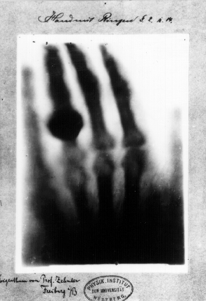
Now that we know what X-rays do when they reach their destination and how they interact with material, the only question that remains is... How is an X-ray produced?

The centre of an X-ray machine is an electrode pair, a cathode and an anode, which is placed inside a glass vacuum tube. The cathode is a heated filament that a current is passed through; the current is what makes it hot. The heat shoots electrons off of the filament surface. The positive anode, a flat surface made of tungsten, pulls the electrons across the glass vacuum tube.
The difference in the voltage between the cathode and anode is extremely high, so the electrons fly through the tube with a huge amount of force and speed. When a speeding electron collides with an atom on the anode, it knocks the electron onto the lower orbital. An electron in the higher level then falls to the one below it, releasing a photon from the extra energy. It's a big fall, so the photon has a high energy level. This high energy photon is an X-ray photon.
There is another way to make a photon without a free electron smashing into another secured electron. An atom's nucleus can pull a high-speed electron enough to change its course, almost like a comet whipping around the sun. The electron decelerates and changes direction as it speeds past the atom. This change in speed causes the electron to emit additional energy in the form of an X-ray photon.
 The free electron crashes with the tungsten atom, which knocks an electron out of a lower orbital. A higher orbital electron fills the empty position, releasing the excess energy as photons
 The free electron is attracted to the incoming tungsten atom nucleus. As the electron passes, the nucleus slightly changes it's course. The electron will lose energy, but it will be released as an X-ray photon.
The entire mechanism is covered by a thick lead barrier. This keeps the X-rays from escaping in all directions. A small cut out in the shield lets some of the X-ray photons escape in a thin beam. The photons escape through a line-up of filters on its way to the patient. A capture device on the other side of the patient records the pattern of light that passes all the way through the person’s body making a image on a common imaging film.
There are two processes that take place during the conversion of the X-ray
The first is bremsstrahlung ( which translates into "breaking rays") and it occurs when the electrons moving at high speed are slowed or put to a full halt by forces of any atom it comes across. The second is the K-shell emission where the once-heavy atom is in the lowest state of energy it can go. Both of these processes are vital in X-ray production although it is often preferred to use the latter over the first.
Clayton Ellis, Donald Lacy, Catherine Little, Heather Mace, Lionel Sandner, Pauline Webb, Otto Wevers, Sandy Wohl.
Investigating Science 10, Canada, Pearson Canada (2009)
Harris, Tom. "How X-rays Work" 26 March 2002. HowStuffWorks.com. <http://science.howstuffworks.com/x-ray.htm> 11 April 2012.
Author Unknown. "Making X-rays" Date unknown. Colorado.edu.
http://www.colorado.edu/physics/2000/xray/making_xrays.html 11 April 2012
Author Unknown. "What is Bremsstrahlung?" Date unkown. ndt-en.org.
Author Unknown. "Advantages of X-ray". Date unknown. medindia.net.
http://www.medindia.net/patients/patientinfo/x-ray_advantages.htm 11 April 2012
Author Unknown. "K-shell emission". Date unknown. Colorado.edu.
http://www.colorado.edu/physics/2000/xray/making_xrays3.html 11 April 2012